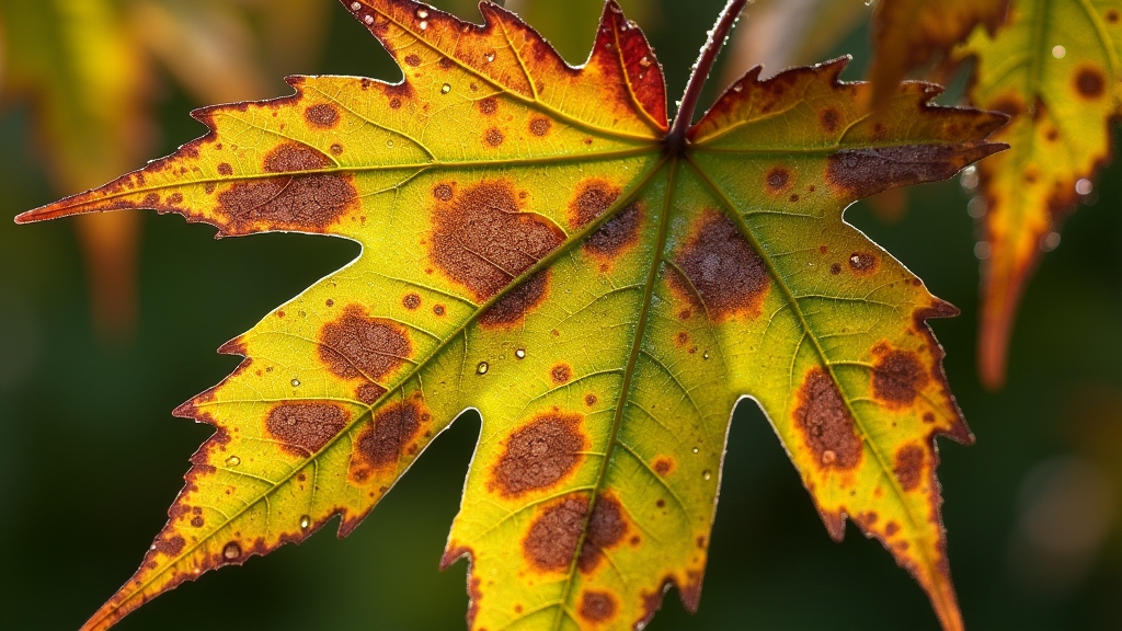
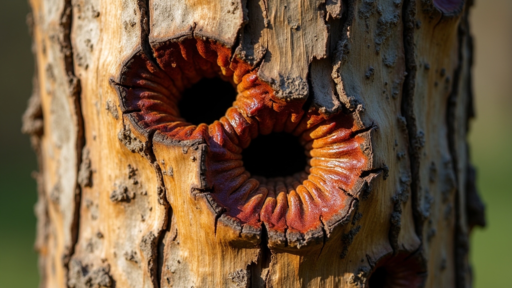
Identify common tree diseases by using a seasonal checklist. Inspect late winter bark for cankers, cracks, and sap bleed. In spring, check uniform budbreak and leaf spots, blotches, or angular water-soaked lesions. Monitor summer wilting, dieback, thinning foliage, and premature fruit drop. Note mushrooms at the root flare, shelf-like conks on trunks, and powdery coatings on leaves. Verify soil moisture and mulch depth, prune infected tissue, and sanitize tools. Call an ISA arborist for rapid decline or structural risk. More follows.
Key Takeaways
- Inspect leaves for discrete spots, blotches, powdery coatings, or angular water-soaked lesions; note black specks (acervuli/pycnidia) indicating fungal fruiting.
- Examine bark and trunk for sunken cankers, bark splits, sap exudation, and frass or entry holes that suggest pathogens or borers.
- Assess canopy for wilting patterns, tip dieback, thinning foliage, and abnormal shoot growth such as water sprouts or epicormic shoots.
- Look for fungal signs: mushrooms at the root flare, shelf-like conks on trunks, and layered perennial brackets indicating internal decay.
- Document season, site conditions, and photos; test soil moisture, confirm mulch depth, and sanitize tools between trees to avoid spread.
Seasonal Checklist: What to Look For Year-Round

When should symptoms raise concern across the seasons?
In late winter, inspect bark and scaffold branches for cankers, sunscald, frost cracks, and oozing exudates; note abnormal sap flow and woodpecker foraging that may indicate borer activity.
Early spring, evaluate budbreak uniformity, flower set density, and dieback at twig tips; flag delayed phenology relative to species norms.
Late spring to early summer, assess shoot elongation, water sprout proliferation, and epicormic growth suggesting stress; probe for soft, discolored cambium around suspect lesions.
Mid–late summer, monitor for wilting unresponsive to adequate soil moisture, resin bleeding, frass at bark fissures, and premature cone or fruit drop.
Early autumn, compare timing of senescence among conspecifics; record abnormal branch mortality patterns and bracket fungi emergence.
Year-round, test soil moisture and drainage, confirm mulch depth, and verify pruning wounds are clean and sealed by callus.
Document findings with dated photos to establish disease progression baselines.
Leaf Symptoms: Spots, Blotches, and Unnatural Color Changes

Building on seasonal surveillance of stems and branches, foliage offers high-resolution indicators of disease activity. Leaf spots typically present as discrete lesions with defined margins; concentric rings suggest anthracnose, while purple halos implicate bacterial leaf spot. Blotches with irregular margins often signal fungal pathogens; necrotic tissue bordered by chlorotic halos indicates active toxin diffusion. Unnatural color changes—interveinal chlorosis, marginal scorch, bronzing—require differentiation from nutrient deficiency and abiotic stress by pattern symmetry, onset timing, and presence of sporulation.
| Symptom pattern | Diagnostic cues and next steps |
| Tiny black specks in lesions | Look for acervuli/pycnidia; confirm with hand lens; prune infected leaves; improve airflow. |
| Angular, water-soaked spots | Bacterial etiology likely; avoid overhead irrigation; sanitize tools; copper only if label permits. |
| Interveinal chlorosis | Test soil/root-zone; rule out iron/manganese deficiency; check pH and compaction. |
| Marginal necrosis after heat | Abiotic scorch; verify irrigation uniformity; mulch; differentiate from leaf blight by lack of sporulation. |
Action: document distribution (sun vs. shade), lesion progression, and host specificity; collect samples early for lab confirmation.
Bark and Trunk Clues: Cankers, Cracks, and Oozing Sap

On inspection of the trunk and limbs, the observer should identify sunken cankers: localized, depressed lesions with cracked margins and discolored cambium.
They should recognize bark splitting by linear fissures aligned with growth stress or frost injury, noting exposed wood and callus formation.
They should note sap exudation (gummosis or bleeding) by sticky, amber or dark ooze that attracts insects and indicates potential pathogen entry.
Identifying Sunken Cankers
How does a sunken canker reveal itself on bark and trunk surfaces? It presents as a distinctly depressed lesion with sharp margins where healthy cambium abuts necrotic tissue.
The bark often appears darker, desiccated, and slightly wrinkled; margins may show a callus ridge attempting compartmentalization.
Probe firmness: diseased bark feels brittle and may slough, exposing cinnamon to dark-brown phloem. Look for elliptical or target-like contours along branch collars or on the trunk where prior wounds existed.
Diagnostic cues include reduced twig vigor distal to the canker, sparse foliage, and dieback progressing above the lesion.
During wet periods, amber to milky exudate or fungal stromata may appear at edges.
Action: sterilize pruners, excise infected wood 10–15 cm beyond visible margins, improve airflow, reduce irrigation splash, and monitor expansion.
Recognizing Bark Splitting
While sunken cankers create localized, depressed lesions, bark splitting manifests as linear to stair-stepped fissures that may run with the grain along trunks or major scaffolds. Observers should differentiate radial frost cracks from longitudinal stress splits by mapping crack orientation, length, and edge character.
Fresh splits display sharply delaminated bark margins and exposed cambium; chronic splits show rolled callus, bark shingle-lifting, and compartmentalization seams. Probe gently to gauge depth without widening the defect.
- 1) Note site factors provoking failure: southwest exposure, freeze–thaw swing, drought–rewet cycles, and recent pruning that shifted load.
- 2) Prioritize safety: inspect for shear planes near attachments, included bark, and movement under light leverage.
- 3) Act: reduce crown leverage, install flexible support where warranted, and sanitize tools between trees.
Noting Sap Exudation
Sap flow tells-tale pathology when it escapes the vascular system at cankers, cracks, or borer wounds. Observers should first locate exudation points, distinguishing pressure-driven bleeding from passive seepage.
Note viscosity, color, and odor: clear, watery flow suggests frost cracks; amber, resinous pitch implicates conifer defense; sour, fermenting flux indicates bacterial wetwood or alcoholic flux.
Examine for frass, entry holes, or galleries to implicate borers. Assess margin tissue: callus formation indicates chronic canker; necrotic rims imply active infection.
Record seasonality and environmental context—spring bleeding after pruning differs from midsummer flux in heat-stressed wood.
Standardize sampling: photograph scale references, swab margins for culture or PCR, and probe for cambial discoloration.
Implement immediate sanitation pruning, tool disinfection, wound size minimization, and vector exclusion where indicated.
Branch and Canopy Issues: Wilting, Dieback, and Thinning Foliage
Although symptoms can overlap across disorders, branch and canopy issues are best parsed by pattern, progression, and location.
Wilting confined to midday that recovers overnight suggests transient water stress; persistent flagging on discrete branches implies vascular obstruction or root impairment.
Dieback typically starts at twig tips and advances proximally; sharp boundaries point to mechanical injury or canker margins, while diffuse decline indicates systemic dysfunction.
Thinning foliage with shortened internodes and undersized leaves signals chronic stress predating the current season.
Immediate actions emphasize verification and triage: assess soil moisture profile, root collar depth, and mulch/compaction; prune back to live wood with visible growth rings; and map affected sectors to track spread.
Prioritize structural safety where dead scaffolds exist.
1) Notice the sudden hush of a canopy losing density—an early alarm.
2) Feel urgency when a single flagging limb foreshadows systemic failure.
3) Respect the line between green and lifeless wood—cut decisively there.
Document dates, weather, irrigation changes, and pruning responses to refine diagnosis.
Fungal Signs: Mushrooms, Conks, and Powdery Growths
Branch and canopy patterns set the stage, but external fungal structures often confirm underlying pathology. Fruiting bodies provide high-certainty cues about infection location and severity. Mushrooms at the root flare or along buttress roots indicate root and butt rot; shelf-like conks on trunks signal advanced heartwood decay; powdery coatings on foliage point to superficial leaf infection. Record host species, distribution, and density to refine identification.
- Mushrooms: clustered at soil-line, short-lived, often following rain; probe for softened wood and basal hollows.
- Conks (brackets): perennial, layered; align with internal decay columns—map their vertical position.
- Powdery growths: white to gray mycelial mats on leaves, shoots; inspect for distorted tissues and reduced vigor.
| Structure | Typical Location | Diagnostic Implication |
| Mushrooms | Root flare, soil-line | Root/butt decay |
| Conks | Trunk, large limbs | Heartwood decay |
| Powdery growth | Leaves, shoots | Epidermal infection |
Clean tools between trees, photograph specimens, and note seasonality to support accurate disease attribution.
When to Act: Differentiating Disease From Pests or Stress and Calling an Arborist
When should intervention begin, and what warrants professional help? Action starts with triage. Distinguish disease from pests or abiotic stress by pattern, timing, and tissue specificity.
Diseases often show progressive wilting, cankers with necrotic margins, vascular discoloration, or sectorial dieback. Insect injury reveals galleries, frass, exit holes, or chewing patterns. Abiotic stress presents uniform symptoms across a canopy edge or exposure side, with no pathogen signs.
Key triggers for calling an ISA Certified Arborist include rapid canopy decline (>20% in a season), structural compromise, or proximity risks. Collect evidence: high‑resolution photos, site history (grading, de‑icing salts, drought), and simple diagnostics—scratch test for cambial viability, pruning cuts to inspect staining, soil moisture and compaction checks.
Call an ISA Certified Arborist when canopy decline accelerates, structure falters, or proximity risks rise. Gather clear evidence.
1) Fear of losing a legacy tree compels prompt assessment—hesitation costs structure.
2) Hope rises when early sanitation pruning halts inoculum spread.
3) Relief arrives as a clear management plan aligns treatment, monitoring, and risk mitigation.
Frequently Asked Questions
How Do Soil Conditions Influence Susceptibility to Tree Diseases?
Soil conditions modulate host vigor and pathogen activity. Compaction, poor drainage, pH imbalance, and nutrient deficiencies predispose trees to root rots and cankers. Prescribe: aeration, organic matter incorporation, drainage correction, pH adjustment, balanced fertilization, and routine soil assays to mitigate susceptibility.
Can Pruning Practices Prevent or Worsen Common Tree Diseases?
Yes. Proper pruning reduces inoculum, improves airflow, and optimizes wound compartmentalization; poor timing and technique amplify infection risk. He schedules dormant-season cuts, sterilizes tools between trees, avoids large flush cuts, preserves branch collars, and applies immediate sanitation removal of symptomatic tissues.
Are Certain Tree Species Naturally Resistant to Prevalent Local Pathogens?
Yes. Species exhibit innate resistance profiles shaped by coevolution. Practitioners should consult regional extension pathogen-resistance matrices, prioritize locally adapted cultivars, diversify genera, verify certified stock, and monitor sentinel plantings, adjusting site selection, spacing, and sanitation to exploit resistance and minimize inoculum pressure.
How Do Climate Change and Extreme Weather Affect Disease Outbreaks?
Climate warming and extremes intensify pathogen survival, vector activity, and host stress, precipitating outbreaks. Practitioners should monitor degree-day thresholds, moisture anomalies, and wounding events; adjust species selection, irrigation, and sanitation; expand scouting post-storm; implement preventative fungicides or pruning timed to phenology and forecasted risk.
What Lab Tests Confirm a Suspected Tree Disease Diagnosis?
Confirmation relies on culture and isolation, PCR/qPCR for pathogen DNA, ELISA for viral/fungal antigens, microscopy of stained tissues, nematode extraction, nutrient and soil assays, and pathogenicity tests (Koch’s postulates). Submit fresh symptomatic tissues; include lesion margins and metadata.
Final Thoughts
Accurate disease ID starts with seasonal observation and pattern recognition. Watch for expanding leaf lesions, active cankers with sap bleed, progressive dieback, and persistent fruiting bodies at the base or trunk—then act quickly with sanitation cuts and clean tools. When decline accelerates or structure is at risk, follow ANSI/ISA best practices and bring in a professional diagnosis and plan.
Need help confirming what you’re seeing or stopping spread fast?
Trust the experts at Tinley Park Tree Service. We provide standards-based pruning, safe removals, and 24/7 emergency service. Learn why hiring a pro matters: Why you should hire a professional tree service.
Take the next step:
- Get a diagnosis & treatment plan: Contact us for a prompt arborist assessment.
- Explore services: Full residential tree care | Stump grinding
- Read more: Blogs on trimming, pruning, and disease prevention.
Keep your trees healthy—and your property safe—with certified care that’s done right the first time.


